Sunday, April 26, 2015

Big Data Meets Quantum Chemistry Approximations: The ∆-Machine Learning Approach

Contributed by +Jan Jensen 
Figure 1. Two hypothetical property profiles connecting two constitutional isomers of C$_7$H_$_{10}$O$_2$. The Δ-model, estimates the difference between baseline and target line properties (arrow) which differ in level of theory (b → t), geometry ($R_b$ → $R_t$), and property ($E_b$ → $H_t$). Reprinted with permission from J. Chem. Theory Comput. 2015, ASAP. Copyright (2015) American Chemical Society.

The idea behind this method is best explained by a specific example.  The G4MP2 enthalpies [$H_t(R_t)$] of  C$_7$H_$_{10}$O$_2$ isomers are estimated using PM7 electronic energies [$E_t(R_b)$] by 
$$H_t(R_t) \approx \Delta_b^t(R_b) = E_b(R_b)+ \sum_{i=1}^N\alpha_i e^{|R_i-R_b|/\sigma}$$
Here {$\alpha_i$} and $\sigma$ are parameters found by regression using a training set of $N$ molecules and $|R_i-R_b|$ is a measure of similarity between the target molecule and training molecule $i$.  The latter is described in more detail here, but I found it pretty interesting so I am summarizing it here.

A Coulomb matrix ($\mathbf{C}$) is constructed for each molecule
$$
C_{kl}= \begin{cases}
 0.5 Z_k^{2.4} & \text{if }  i=j\\
 Z_kZ_l/r_{kl}& \text{if } i \ne j
\end{cases}
$$
where $r_{kl}$ is the distance between atom $k$ and $l$ and $Z_k$ is the nuclear charge of atom $k$. Then the elements are sorted such that the diagonal elements are in descending order and the similarity is computed by
$$|R_i-R_b| = \sum_{k,l} |C_{kl}^i - C_{kl}^b | $$
Using this approach and a training set of ($N$ =) 1000 molecules the G4MP2 atomization enthalpies of 6095 constitutional isomers of C$_7$H_$_{10}$O$_2$ can be reproduced with a MAE of 3.9 kcal/mol using PM7, compared to an MAE of 6.4 kcal/mol for uncorrected PM7.  Using PBE or B3LYP/6-31G(2df,p) the MAE can be brought below 1 kcal/mol using a 1K training set.

In another interesting application the MAE of RHF/6-31G(d) relative to CCSD(T)/6-31G(d) atomization energies for the same set of molecules can be reduced from 3 to less than 1 kcal/mol using a 1K training set.

This is thus a very interesting approach for obtaining chemical accuracy using methods that are sufficiently fast to study thousands of molecules. The caveat is that about 1000 high level calculations appears to be needed to train the method but perhaps more generally applicable parameter sets can be found using, for example, functional group identification.


This work is licensed under a Creative Commons Attribution 4.0  

Wednesday, April 22, 2015

Electron-Driven Proton Transfer Along H2O Wires Enables Photorelaxation of πσ* States in Chromophore−Water Clusters

More than just shifting state energies, polar solvents may actively participate in the photochemistry of excited molecules


Szabla, R.; Šponer, J.; Góra, R. W. J. Phys. Chem. Lett. 2015, 6, 1467-1471.

Highlighted by Mario Barbatti

For decades, it has been well known that solvents, especially polar ones, have a large impact on the photodynamics of chromophores. Phenomenological models, such as the Lim Proximity effect (1), for instance, have been developed to describe how the energy shift caused by the solvent molecules determines radiative and non-radiative rates.

These early approaches focused mostly on the description of the shape of the potential energy surfaces and on the relative shift between them. From that tradition, we learned, for example, the important heuristic rule telling that, in comparison to the gas phase, water stabilizes the ππ* state of the chromophore, while it destabilizes the nπ* state. Those models, however, did not consider that new electronic states arising from the chromophore-solvent interaction could play a major role in the fate of the excited system.

From the photochemical point of view, the solvent was understood as an important but passive factor. Many simulations relied on this important-but-passive hypothesis, to restrict, for instance, the quantum-mechanical region in QM/MM modelling to the chromophore only, saving precious computational time.

In the last years, however, the important-but-passive hypothesis has been challenged by a number case studies (2). Diverse computational simulations have shown that the solvent may indeed play an active role in photochemistry. (I have myself contributed to the field by showing a case where a water-to-chromophore electron transfer could create a conical intersection (3).)

The paper by Szabla, Šponer, and Góra (4) belongs to this new tradition, the important-and-active hypothesis.

Using surface hopping simulations based on ADC(2) excited states, they investigated the ultrafast dynamics of 2-aminooxazole (AMOX) microsolvated by water. They found out that an important radiationless pathway for the AMOX-(H2O)5 cluster involves an electron-driven proton transfer along water wires. This process occurs in an electronic state characterized by an electron transfer from an n orbital at the chromophore to a σ* orbital in one of the water molecules (Fig. 1).

Fig. 1 - Electron-driven proton transfer along water wires.
Szabla, Šponer, and Góra note that similar deactivation mechanism has been observed before in simulations of other heterocyclic chromophores. This means that it may be a common pattern in the photochemistry of these compounds.

Although this is is matter for speculation, all these examples imply that we cannot restrict ourselves to credit polar solvents a passive role only. Any new investigation, either experimental or theoretical, has now to take into account the possibility that the solvent may actively be contributing to the photochemistry.

In particular, for the next generation of excited-state QM/MM simulations, the message (and the cost) is clear: quantum-mechanical microsolvation is simply mandatory.

References
(1) Lim, E. C. Proximity effect in molecular photophysics: dynamical consequences of pseudo-Jahn-Teller interaction. J. Phys. Chem. 1986, 90, 6770-6777. doi: 10.1021/j100284a012

(2) Liu, X.; Sobolewski, A. L.; Borrelli, R.; Domcke, W. Computational investigation of the photoinduced homolytic dissociation of water in the pyridine-water complex. Phys. Chem. Chem. Phys. 2013, 15, 5957-5966. doi: 10.1039/C3CP44585B

(3) Barbatti, M. Photorelaxation Induced by Water–Chromophore Electron Transfer. J. Am. Chem. Soc. 2014, 136, 10246-10249. doi: 10.1021/ja505387c

(4) Szabla, R.; Šponer, J.; Góra, R. W. Electron-Driven Proton Transfer Along H2O Wires Enables Photorelaxation of πσ* States in Chromophore–Water Clusters. J. Phys. Chem. Lett. 2015, 6, 1467-1471. doi: 10.1021/acs.jpclett.5b00261

Wednesday, April 15, 2015

The Fluorenyl Cation

Costa, P.; Trosien, I.; Fernandez-Oliva, M.; Sanchez-Garcia, E.; Sander, W. Angew. Chem. Int. Ed. 2015, 54, 2656-2660
Contributed by Steven Bachrach.
Reposted from Computational Organic Chemistry with permission

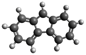
Is the fluorenyl cation 1 antiaromatic or non-aromatic? This is still an open question. But the recent paper by Costa, et al. provides a new path towards potentially answering this question; they have finally synthesized this molecule.1

By photolizing 2 in low-density amorphous ice (LDA ice) and in deuterated ice at 8 K, they have identified a new IR spectrum.

To identify the origin of these spectra, they optimized the geometry of the fluorenyl cation 1 at B3LYP-D3/def2-TZVP (see Figure 1) and computed its IR spectra. These computed IR frequencies were then scaled by 0.97. The agreement between the computed and experimental frequencies is quite reasonable, and the isotopic shifts are also reasonably well reproduced. The agreement is not perfect, as seen in Table 1. Hopefully, further experiments will now be carried out to try to answer the lead question of this post.

Figure 1. B3LYP-D3/def2-TZVP optimized geometry of 1.
Table 1. Experimental and computed IR frequencies (cm-1)
and isotopic shift (in parentheses) of 1.
Calc.
Exp.
1008.8
986
1106.8 (+2.1)
1076.8 (+1.7)
1152.8
1117.2
1198.6
1163.5
1267.0
1235.1
1373.4 (-16.4)
1343.7
1510.8(-8.3)
1469.0 (-7.3)
1530.7 (-3.2)
1490.5 (-1.0)
1616.8(-6.4)
1575.7(-4.4)
1640.9 (0.0)
1601.2 (-4.0)

References

(1) Costa, P.; Trosien, I.; Fernandez-Oliva, M.; Sanchez-Garcia, E.; Sander, W. "The Fluorenyl Cation,"Angew. Chem. Int. Ed. 201554, 2656-2660, DOI: 10.1002/anie.201411234.

InChIs

1: InChI=1S/C13H9/c1-3-7-12-10(5-1)9-11-6-2-4-8-13(11)12/h1-9H/q+1
InChIKey=KZCNYQVQQBONEY-UHFFFAOYSA-N


This work is licensed under a Creative Commons Attribution-NoDerivs 3.0 Unported License.

Tuesday, April 7, 2015

The Nucleoside Uridine Isolated in the Gas Phase

Peña, I.; Cabezas, C.; Alonso, J. L. Angew. Chem. Int. Ed. 2015, 54, 2991-2994
Contributed by Steven Bachrach.
Reposted from Computational Organic Chemistry with permission

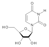
To advance our understanding of why ribose takes on the furanose form, rather than the pyranose form, in RNA, Alonso and co-workers have examined the structure of uridine 1 in the gas phase.1

1
Uridine is sensitive to temperature, and so the laser-ablation method long used by the Alonso group is ideal for examining uridine. The microwave spectrum is quite complicated due to the presence of many photofragments. Careful analysis lead to the identification of a number of lines and hyperfine structure that could be definitively assigned to uridine, leading to experimental values of the rotational constants and the diagonal elements of the 14N nuclear quadrupole coupling tensor for each nitrogen. These values are listed in Table 1.

Table 1. Experimental and calculated rotational constants (MHz), quadrupole coupling constants (MHz) and relative energy (kcal mol-1).


calculated


Expt.
anti/C2’-endo-g+
syn/C2’-endo-g+
anti/C3’-endo-g+
anti/C2’-endo-t
syn/C3’-endo-g+
A
885.98961
901.2
935.8
790.0
799.7
925.5
B
335.59622
340.6
308.4
352.6
330.6
300.4
C
270.11210
276.6
266.6
261.4
262.9
264.0
14N1χxx
1.540
1.50
1.82
1.48
1.46
1.82
14N1χyy
1.456
1.43
0.73
1.71
1.81
-0.72
14N1χzz
-2.996
-2.93
-2.56
-3.19
-3.27
-1.11
14N3χxx
1.719
1.74
2.03
1.78
1.62
1.98
14N3χyy
1.261
1.11
0.47
1.34
1.51
-0.75
14N3χzz
-2.979
-2.85
-2.50
-3.12
-3.13
-1.23
Rel E

0.0
1.10
1.90
2.00
2.15

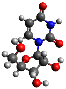
In order to assign a 3-D structure to these experimental values, they examined the PES of uridine with molecular mechanics and semi-empirical methods, before reoptimizing the structure of the lowest 5 energy structures at MP2/6-311++G(d,p). Then, comparison of the resulting rotational constants and 14N nuclear quadrupole coupling constants of these computed structures (see Table 1) led to identification of the lowest energy structure (anti/C2’-endo-g+, see Figure 1) in best agreement with the experiment.Once again, the Alonso group has demonstrated the value of the synergy between experiment and computation in structure identification.

Figure 1. MP2/6-311++G(d,p) optimized structure of 1 (anti/C2’-endo-g+).


References

(1) Peña, I.; Cabezas, C.; Alonso, J. L. "The Nucleoside Uridine Isolated in the Gas Phase," Angew. Chem. Int. Ed. 201554, 2991-2994, DOI: 10.1002/anie.201412460.


Inchis:

1: Inchi=1S/C9H12N2O6/c12-3-4-6(14)7(15)8(17-4)11-2-1-5(13)10-9(11)16/h1-2,4,6-8,12,14-15H,3H2,(H,10,13,16)/t4-,6-,7-,8-/m1/s1
InChiKey=DRTQHJPVMGBUCF-XVFCMESISA-N


This work is licensed under a Creative Commons Attribution-NoDerivs 3.0 Unported License.