Wednesday, December 28, 2016

Ultra-fast computation of electronic spectra for large systems by tight-binding based simplified Tamm-Dancoff approximation (sTDA-xTB)

Stefan Grimme and Christoph Bannwarth (2016)
Contributed by Jan Jensen


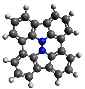
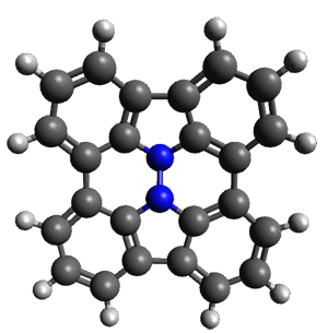
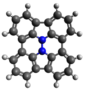
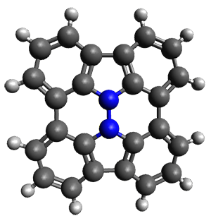
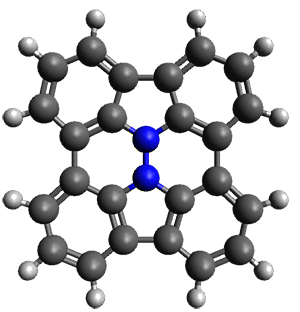
Grimme and Bannwarth presents the xTB method, a modified DFTB2 (SCC-DFTB) method combined with Grimme's simplified Tamm-Dancoff  approximated (sTDA) TD-DFT procedure for the rapid calculations of electronic spectra.  The two main modifications are 1) the addition of diffuse basis functions for some elements to accurately describe Rydberg states and 2) the SCF is replaced by a single diagonalization and the atomic-charge dependent term is evaluated only once using specifically designed charges. The method is parameterized against CC and DFT calculations for all elements up to Zn and more elements are in the works.

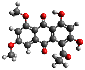
SCS-CC2/aug-cc-VXZ (X = T or D depending on molecule size) vertical singlet-singlet excitation energies are reproduced with a MAE of 0.27 eV.  For comparison, the corresponding MAEs for TD-PBE/def2-TZVP and TD-PBE0/def2-SV(P) are 0.67 and 0.31 eV.  The method also yields reasonably accurate ECD spectra.  

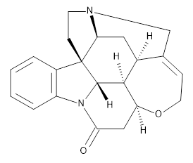
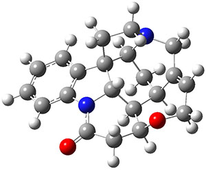
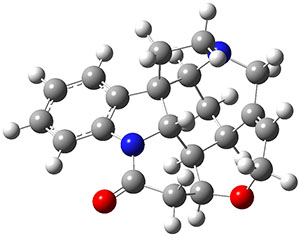
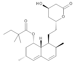
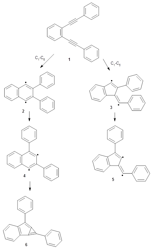
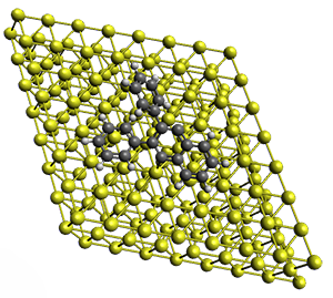
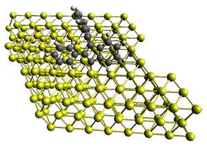
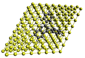
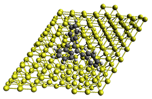
The method is quite fast requiring only a few minutes of CPU time for molecules with 500-1000 atoms. A very recent paper uses an as-yet-unpublished variant called GFN-xTB to perform an single point calculation on a P450 enzyme (7,500 atoms) without recourse to fragmentation.


This work is licensed under a Creative Commons Attribution 4.0

Friday, December 16, 2016

Evidence of a Nitrene Tunneling Reaction: Spontaneous Rearrangement of 2-Formyl Phenylnitrene to an Imino Ketene in Low-Temperature Matrixes

Nunes, C. M.; Knezz, S. N.; Reva, I.; Fausto, R.; McMahon, R. J.,  J. Am. Chem. Soc. 2016, 138, 15287-15290
Contributed by Steven Bachrach
Reposted from Computational Organic Chemistry with permission

Reva and McMahon report a very nice experimental and computational study implicating hydrogen atom tunneling in the rearrangement of the nitrene 1 into the ketene 2.1 The reaction is carried out by placing azide 3 in an argon matrix and photolyzing it. The IR shows that at first a new compound A is formed and that over time the absorptions of A erode and those of a second compound B grow in. This occurs whether the photolysis continues or not over time.


IR spectra were computed at B3LYP/6-311++G(d,p) for compounds 31 and 2 and they match up very well with the recorded spectra of A and B, respectively. The triplet state of nitrenes are typically about 20 kcal mol-1 lower in energy than the singlet states. The EPR spectrum confirms that 1 is a triplet.
So how does the conversion of 31 into 2 take place, especially at 10 K? The rate constant for this conversion at 10 K is estimated as 1 x 10⁻⁵ s-1, which implies a barrier from classical transition state theory of only 0.2 kcal mol-1. That low a barrier seems preposterous, and suggests that the reaction may proceed via tunneling. This notion is supported by the experiment on the deuterated analogue, which shows no conversion of 1D into 2D.

The authors propose that 31 undergoes a hydrogen migration on the triplet surface through transition state 34 to give 32, which then undergoes intersystem crossing to give singlet 2. The structures of these critical points calculated at B3LYP/6-311++G(d,p) are shown in Figure 1. The computed activation barrier is 20.7 kcal mol-1. (The barrier height ranges from 16.7 to 23.0 with a variety of different computational methods.) This large barrier precludes a classical over-the-top reaction and points towards tunneling. The barrier width is estimated at about 2.1 Å. WKB computations estimate the tunneling half time of about 21 min, somewhat smaller than in the experiments, and the estimate for the deuterated species is 150,000 years.

31

34

32
Figure 1. B3LYP/6-311++G(d,p) optimized structures of 3132, and the TS 34.


References

1) Nunes, C. M.; Knezz, S. N.; Reva, I.; Fausto, R.; McMahon, R. J., "Evidence of a Nitrene Tunneling Reaction: Spontaneous Rearrangement of 2-Formyl Phenylnitrene to an Imino Ketene in Low-Temperature Matrixes." J. Am. Chem. Soc. 2016, 138, 15287-15290, DOI: 10.1021/jacs.6b07368.


InChIs:

1: InChI=1S/C7H5NO/c8-7-4-2-1-3-6(7)5-9/h1-5H
InChIKey=QZTZBORTPUZAGF-UHFFFAOYSA-N
2: InChI=1S/C7H5NO/c8-7-4-2-1-3-6(7)5-9/h1-4,8H
InChIKey=ZWHBMBVIYUVTGT-UHFFFAOYSA-N


'
This work is licensed under a Creative Commons Attribution-NoDerivs 3.0 Unported License.

Wednesday, November 30, 2016

ANI-1: An extensible neural network potential with DFT accuracy at force field computational cost

Justin S. Smith, Olexandr Isayev, Adrian E. Roitberg (2016)
Contributed by Jan Jensen


This paper basically presents a neural network force field, which the authors call a neural network potential (NNP).  The authors heavily modify the Behler-Parinello symmetry functions (also used in this CCH) to improve the transferability and train it against 13.8 million ωB97X/6-31G(d) energies computed for CHON-containing molecules with 8 or less non-hydrogen atoms. This huge training set made it possible to parameterise a neural net with three hidden layers with a total of 320 nodes and 124,033 optimisable parameters.  Deep learning indeed.  

What makes this work particularly exiting is that the NNP appears to be transferable to larger molecules. For example, the figure above shows that the NNP can reproduce the relative ωB97X/6-31G(d) energies of retinol conformers with en RMSE of 0.6 kcal/mol.  For comparison the corresponding value for DFTB (not clear if it's DFTB2 or DFTB3) is 1.2 kcal/mol, although ωB97X/6-31G(d) is not the definitive reference by which to judge DFTB accuracy.

I think this work holds a lot of promise. One of the key challenges is to reduce the size of the training set to a point where high level calculations can be used to compute the energies. Alternatively, perhaps approaches like ∆-machine learning can be used to correct the NNP using a smaller representative training set.


This work is licensed under a Creative Commons Attribution 4.0

Wednesday, November 16, 2016

Calculation of NMR Spin–Spin Coupling Constants in Strychnine

Helgaker, T.; Jaszuński, M.; Świder, P. J. Org. Chem. 2016
Contributed by Steven Bachrach
Reposted from Computational Organic Chemistry with permission

Helgaker, Jaszunski, and Swider1 have examined the use of B3LYP with four different basis sets to compute the spin-spin coupling constants in strychnine 1.

1
They used previously optimized coordinates of the two major conformations of strychnine, shown in Figure 1.

Conformer A

Conformer B
Figure 1. Confrmations of strychnine 1.

They tested four basis sets designed for NMR computations: pcJ-0,2 pcJ-1,2 6-31G-J,3 and 6-311G-J.3 pCJ-0 and 6-31G-J are relatively small basis sets, while the other two are considerably larger.

All four basis sets provide values of the 122 J(C-H) with a root mean square deviation of less than 0.6 Hz. J(HH) and J(CC) coupling constants are also well predicted, especially with the larger pcJ-1 basis set. They also examined the four Ramsey terms in the coupling model. The Fermi contact term dominates, and if the large pcJ-1 basis set is used to calculate it, and the smaller pcJ-0 basis set is used for the other three terms, the RMS error only increases from 0.18 to 0.20 Hz. Taking this to the extreme, they omitted calculating any of the non-Fermi contact terms, with again only small increases in the RMS – even with the small pcJ-0 basis set. Considering the computational costs, one should seriously consider whether the non-Fermi contact terms and a small basis set might be satisfactory for your own problem(s) at hand.

References

1) Helgaker, T.; Jaszuński, M.; Świder, P., "Calculation of NMR Spin–Spin Coupling Constants in Strychnine." J. Org. Chem. 2016, ASAP, DOI: 10.1021/acs.joc.6b02157.
2) Jensen, F., "The Basis Set Convergence of Spin−Spin Coupling Constants Calculated by Density Functional Methods." J. Chem. Theor. Comput. 2006, 2, 1360-1369, DOI: 10.1021/ct600166u.
3) Kjær, H.; Sauer, S. P. A., "Pople Style Basis Sets for the Calculation of NMR Spin–Spin Coupling Constants: the 6-31G-J and 6-311G-J Basis Sets." J. Chem. Theor. Comput. 2011, 7, 4070-4076, DOI: 10.1021/ct200546q.

InChIs

Strychnine 1: InChI=1S/C21H22N2O2/c24-18-10-16-19-13-9-17-21(6-7-22(17)11-12(13)5-8-25-16)14-3-1-2-4-15(14)23(18)20(19)21/h1-5,13,16-17,19-20H,6-11H2/t13-,16-,17-,19-,20-,21+/m0/s1
InChIKey=QMGVPVSNSZLJIA-FVWCLLPLSA-N

'
This work is licensed under a Creative Commons Attribution-NoDerivs 3.0 Unported License.

Sunday, October 30, 2016

Automatic chemical design using a data-driven continuous representation of molecules


Rafael Gómez-Bombarelli, David Duvenaud, José Miguel Hernández-Lobato, Jorge Aguilera-Iparraguirre, Timothy D. Hirzel, Ryan P. Adams, and Alán Aspuru-Guzik (2016)
Contributed by Jan Jensen


Chemical space is discrete which makes it hard to search with standard techniques such as gradient-based minimisation.  This paper used a standard machine learning tool called an autoencoder to help solve that problem.  One way to think of an autoencoder is as a data-compressor where one neural network is trained to describe a data set such as an image in some compressed representation and another network is trained to recover the image from the compressed format.

The interesting thing in the context of chemical space is that the compressed format can be a continuous function such as a real-valued vector (latent space). (Another use of autoencoders is dimensionality reduction for data visualization, e.g. as an alternative to principal component analysis.)  This latent space is therefore a continuous representation of the chemical space (a set of SMILES strings) that the autoencoder was trained on.  Another neural net can then be trained to map some chemical property, such as logP values, on this latent space and the space can be searched for regions with desired logP values with techniques as simple as interpolation.

One problem with autoencoders is that they are "lossy" which in this case translates to the fact that not all points in latent space can be decoded to a valid molecule (SMILES string) but the failure rate is relatively low for the two proof-of-concept applications in the paper.

This is a very interesting new tool in the hunt for molecules with new properties.


This work is licensed under a Creative Commons Attribution 4.0

Wednesday, October 26, 2016

More examples of structure determination with computed NMR chemical shifts

Nguyen, Q. N. N.; Tantillo, D. J. “Using quantum chemical computations of NMR chemical shifts to assign relative configurations of terpenes from an engineered Streptomyces host,” J. Antibiotics 2016, 69, 534–540
Khokhar, S.; Pierens, G. K.; Hooper, J. N. A.; Ekins, M. G.; Feng, Y.; Rohan A. Davis, R. A. “Rhodocomatulin-Type Anthraquinones from the Australian Marine Invertebrates Clathria hirsuta and Comatula rotalaria,” J. Nat. Prod., 2016, 79, 946–953
Contributed by Steven Bachrach
Reposted from Computational Organic Chemistry with permission

Use of computed NMR chemical shifts in structure determination is really growing fast. Presented here are a couple of recent examples.

Nguyen and Tantillo used computed chemical shifts with the DP4 analysis to identify the structure of three terpenes 1-3.1 They optimized the geometries of all of the diastereomers of each compound, along with multiple conformations of each diastereomer, at B3LYP/6-31+G(d,p) and then computed the chemical shifts at SMD(CHCl3)–mPW1PW91/6-311+G(2d,p). The chemical shifts were Boltzmann weighted including all conformations within 3 kcal mol-1 of the lowest energy structure.


For 1, the DP4 analysis using just the proton shifts predicted a different isomer than using the carbon shifts, but when combined, DP4 predicted the structure, with 98.8% confidence, shown in the scheme above, and in Figure 1. For 2, the combined proton and carbon shift analysis with DP4 indicated a 100% confidence of the structure shown in the scheme and Figure 1. Lastly, for 3, which is more complicated due to the conformations of the 9-member ring, DP4 predicts with 100% confidence the structure shown in the scheme and Figure 1.

1

2

3
Figure 1. Optimized geometries of 1-3.

Feng, Davis and coworkers have examined a series of anthroquionones from Australian marine sponges.2The structure of one compound was a choice of two options: 4 or 5. Initial geometries were obtain by molecular mechanics and the low energy isomers were then reoptimized at B3LYP/6-31+G(d,p). The chemical shifts were computed using PCM/MPW1PW91/6-311+G(2d,p). Application of the DP4 method indicate the structure to be 4 with a 100% confidence level. The lowest energy conformer of 4 is shown in Figure 2.


Figure 2. Optimized geometry of 4.

References

1) Nguyen, Q. N. N.; Tantillo, D. J. “Using quantum chemical computations of NMR chemical shifts to assign relative configurations of terpenes from an engineered Streptomyces host,” J. Antibiotics 201669, 534–540, DOI: 10.1038/ja.2016.51.
2) Khokhar, S.; Pierens, G. K.; Hooper, J. N. A.; Ekins, M. G.; Feng, Y.; Rohan A. Davis, R. A. “Rhodocomatulin-Type Anthraquinones from the Australian Marine Invertebrates Clathria hirsuta andComatula rotalaria,” J. Nat. Prod., 2016, 79, 946–953, DOI: 10.1021/acs.jnatprod.5b01029.

InChIs

1: InChI=1S/C15H24/c1-10-5-6-15(4)8-11-7-14(2,3)9-12(11)13(10)15/h9-11,13H,5-8H2,1-4H3/t10-,11+,13-,15+/m1/s1
InChIKey=KVSCZIPUFBVHBM-OICBVUGWSA-N
2: InChI=1S/C15H24/c1-10-5-6-15(4)8-11-7-14(2,3)9-12(11)13(10)15/h5,11-13H,6-9H2,1-4H3/t11-,12-,13+,15-/m0/s1
InChIKey=ZLYGJLHCPYVGDA-XPCVCDNBSA-N
3: InChI=1S/C20H32/c1-14-6-9-18-19(3,4)10-11-20(18,5)13-17-15(2)7-8-16(17)12-14/h6,13,15-16,18H,7-12H2,1-5H3/b14-6-,17-13-/t15-,16-,18-,20+/m0/s1
InChIKey=JZGOFJIAHJJJDK-ICZJPRMTSA-N
4: InChI=1S/C18H14O7/c1-7(19)13-10(20)6-11(21)15-16(13)17(22)9-4-8(24-2)5-12(25-3)14(9)18(15)23/h4-6,20-21H,1-3H3
InChIKey=MPQMZEXRJVMYBT-UHFFFAOYSA-N
5: InChI=1S/C18H14O7/c1-7(19)13-10(20)6-11(21)15-16(13)14-9(17(22)18(15)23)4-8(24-2)5-12(14)25-3/h4-6,20-21H,1-3H3
InChIKey=WIKIUXNPFURKNF-UHFFFAOYSA-N

'
This work is licensed under a Creative Commons Attribution-NoDerivs 3.0 Unported License.

Wednesday, October 12, 2016

Expanding DP4: application to drug compounds and automation

Ermanis, K.; Parkes, K. E. B.; Agback, T.; Goodman, J. M. Org. Biomol. Chem., 2016, 14, 3943-3949
Contributed by Steven Bachrach
Reposted from Computational Organic Chemistry with permission

Computational chemistry has had a remarkable impact on the field of structure determination by NMR spectroscopy. The ability to efficiently compute 13C and 1H chemical shifts allows for comparison of the computed chemical shifts of potential structures against the experimental values, a tremendous aid in structure determination (see some examples in previous posts). Goodman and Smith developed the DP4 method1 (see this post) to assist in identifying proper structures by means of statistical distribution of errors and Bayes Theorem.

The Goodman group now reports on workflow solutions to structure prediction using DP4.2 They explore the use of open source computational tools both for predicting conformations and for computing the chemical shifts. They use a set of 10 drugs to test the performance. In general, the original DP4 method works very well in predicting drug structure, despite the fact that DP4 parameters were developed for natural products. The only failure is for simvastatin, where the large number of diastereomers and conformational flexibility prove to be too complex. The open source tools perform just slightly less effectively than the commercial packages, but are certainly a viable route for those with limited resources. The authors also provide a series of python scripts that allow users to create a seamless workflow; these should prove most helpful to the structure determination community.


Simvastatin

References

1) Smith, S. G.; Goodman, J. M. "Assigning Stereochemistry to Single Diastereoisomers by GIAO
NMR Calculation: The DP4 Probability," J. Am. Chem. Soc. 2010132, 12946-12959, DOI:10.1021/ja105035r.
2) Ermanis, K.; Parkes, K. E. B.; Agback, T.; Goodman, J. M. “Expanding DP4: application to drug compounds and automation,” Org. Biomol. Chem.201614, 3943-3949, DOI: 10.1039/c6ob00015k.

InChIs

Simvastatin: InChI=1S/C25H38O5/c1-6-25(4,5)24(28)30-21-12-15(2)11-17-8-7-16(3)20(23(17)21)10-9-19-13-18(26)14-22(27)29-19/h7-8,11,15-16,18-21,23,26H,6,9-10,12-14H2,1-5H3/t15-,16-,18+,19+,20-,21-,23-/m0/s1
InChIKey=RYMZZMVNJRMUDD-HGQWONQESA-N


'
This work is licensed under a Creative Commons Attribution-NoDerivs 3.0 Unported License.

Thursday, September 29, 2016

Multiscale Quantum Mechanics/Molecular Mechanics Simulations with Neural Networks

Lin Shen, Jingheng Wu, and Weitao Yang (2016)
Contributed by Jan Jensen


There has been a lot of work on estimating high level energies from low level energies, most of which have focussed some kind of interpolation or extrapolation.  However, this has been particularly challenging for QM/MM-MD studies where thousands of semiempirical energies contribute to the PMF but only hundreds of high level calculations are feasible. 

Yang and co-workers offer an interesting solution to this problem by using the high level calculations to train a neutral network to estimate the energy correction, which can then be used to estimate the high level energies for all thousands of geometries from the semiempirical QM/MM-MD.  Thus, the PMF can be computed "from scratch" at the high level rather than correcting the low level PMF.

The approach is based on work by Behler and Parrinello, but with three tweaks geared towards this particular problem.

1. The neural network is trained to reproduce an energy correction rather than a total energy.

2. Mulliken charges are used as input, in addition to atomic coordinates of the QM region, to provide some information about the interaction with the MM environment.

3. A subnet is added to the neutral net that depends on the reaction coordinate and the potential of mean force obtained at the low level to improve accuracy.  

The method is tested for three relatively small systems in water, so that high level PMFs can be computed rigorously for comparison. The results are quite impressive. For example, the free energy of glycine zwiterion is ca 8 kcal/mol higher in energy than the neutral form at the SCC-DFT/MM level but ca 8 kcal/mol lower in free energy at the B3LYP/6-31G(d)//MM level of theory. This latter value is reproduced to within 0.1 kcal/mol by the machine learning approach using only 500 B3LYP/6-31G(d)//MM energies to train the neural net. For comparison, the PMF requires 50,000 energy evaluations.


This work is licensed under a Creative Commons Attribution 4.0

Wednesday, September 28, 2016

Polytriangulane

Allen, W. D.; Quanz, H.; Schreiner, P. R. J. Chem. Theory Comput. 2016, 12, 4707–4716
Contributed by Steven Bachrach
Reposted from Computational Organic Chemistry with permission

Cyclopropyl rings can be joined together in a spiro fashion to form triangulanes. An interesting topology can be made by joining the rings to form a helical pattern, as shown in the [9]triangulane 1 below. Allen, Quanz, and Schreiner1 have examined the notion of an infinite helical molecule formed in this way.

1
First, they describe how one can generate the coordinates of such a beast using a closed analytical expression, which is a really nice demonstration of applied geometry. Next, they compute the geometry of a series of [n]triangulanes at M06-2x/6-31G(d). The geometries of [9]triangulane and their largest example, [42]triangulane 2 are shown in Figure 1.

1

2
Figure 1. M06-2x/6-31G(d) optimized geometries of 1 and 2.

They show that the geometry of 2 exhibits a structure that has two different C-C distances: one between the spiro carbons, and the second between the spiro carbon and the methylene carbon. The distance between the spiro carbons is rather short (1.458 Å), suggesting that the bonding here is between carbons that are nearly sp2-hybridized.

Lastly, they discuss the thermodynamics of polytriangulane. They employ a series of homodesmotic reactions to attempt to determine the enthalpy for adding another cyclopropyl ring to an extended triangulane. Unfortunately, the computed enthalpy is quite dependent on functional used. Similar attempts to define the strain energy is also flawed in this way. However, regardless of the functional the enthalpy for adding a cyclopropane ring appears to reach an asymptote rather quickly. So, using [3]triangulane they estimate that the strain energy per mole of cyclopropane in triangulane is about 42.7 kcal mol-1, or about 14 kcal mol-1 of strain due to the spiroannulation.

References

(1) Allen, W. D.; Quanz, H.; Schreiner, P. R. “Polytriangulane,” J. Chem. Theory Comput. 201612, 4707–4716, DOI: 10.1021/acs.jctc.6b00669.

InChIs

1: InChI=1S/C19H22/c1-2-12(1)5-14(12)7-16(14)9-18(16)11-19(18)10-17(19)8-15(17)6-13(15)3-4-13/h1-11H2/t14-,15-,16-,17-,18-,19-/m0/s1
InChIKey=XBTZCZDSKVTALB-DYKIIFRCSA-N


'
This work is licensed under a Creative Commons Attribution-NoDerivs 3.0 Unported License.

Wednesday, September 21, 2016

Enediyne Cyclization on Au(111)

de Oteyza, D. G.; Paz, A. P.; Chen, Y.-C.; Pedramrazi, Z.; Riss, A.; Wickenburg, S.; Tsai, H.-Z.; Fischer, F. R.; Crommei, M. F.; Rubio, A. J. Amer. Chem. Soc. 2016, 138, 10963–10967
Contributed by Steven Bacharach
Reposted from Computational Organic Chemistry with permission

The Bergman cyclization and some competitive reactions are discussed in detail in Chapter 4 of by book. The Bergman cyclization makes the C1-C6 bond from an enediyne. Another, but rarer, option is to make the C1-C5 bond, the Schreiner-Pascal cyclization pathway. de Oteyza and coworkers have examined the competition between these two pathways for 1 on a gold surface, and used STM and computations to identify the reaction pathway.1

The two pathways are shown below. The STM images identify 1 as the reactant on the gold surface and the product is 6. No other product is observed.
Projector augmented wave (PAW) pseudo-potential computations using the PBE functional were performed for the reaction on a Au (111) surface was modeled by a 7 x 7 x 3 supercell. The optimized geometries of the critical points are show in Figure 1.

1

TS(1→2)

TS(1→3)

2

3

TS(2→6)

TS(3→5)

6

5
Figure 1. Optimized geometries of the critical points on the two reaction pathways.

Explicit values of the relative energies are not given in either the paper or the supporting information, but rather a plot shows the relative positions of the critical points. The important points are the following: (a) the barrier for the C1-C5 cyclization is lower than the barrier for the C1-C6 cyclization and 3 is lower in energy than 2; (b) 5 is lower in energy than 6; and (c) the barrier for taking 2 to 6 is significantly below the barrier taking 3 into 5. The barrier for the phenyl migration taking 3 into 5 is so high because of a strong interaction between the carbon radical and a gold atom of the surface. The authors suggest that the two initial cyclizations are reversible, but the very high barrier for forming 5 precludes it from taking place, leaving only the route to 6 as a viable pathway.


References

(1) de Oteyza, D. G.; Paz, A. P.; Chen, Y.-C.; Pedramrazi, Z.; Riss, A.; Wickenburg, S.; Tsai, H.-Z.; Fischer, F. R.; Crommei, M. F.; Rubio, A. “Enediyne Cyclization on Au(111),” J. Amer. Chem. Soc. 2016138, 10963–10967, DOI: 10.1021/jacs.6b05203.


InChIs

1: InChI=1S/C22H14/c1-3-9-19(10-4-1)15-17-21-13-7-8-14-22(21)18-16-20-11-5-2-6-12-20/h1-14H
InChIKey=XOJSMLDMLXWRMT-UHFFFAOYSA-N
2: InChI=1S/C22H14/c1-3-9-17(10-4-1)21-15-19-13-7-8-14-20(19)16-22(21)18-11-5-2-6-12-18/h1-14H
InChIKey=DAUFPUDTOKPCMX-UHFFFAOYSA-N
3: InChI=1S/C22H14/c1-3-9-17(10-4-1)15-22-20-14-8-7-13-19(20)16-21(22)18-11-5-2-6-12-18/h1-14H
InChiKey=>FYBPBPGPMCJQNF-UHFFFAOYSA-N
4: InChI=1S/C22H14/c1-3-9-17(10-4-1)20-15-19-13-7-8-14-21(19)22(16-20)18-11-5-2-6-12-18/h1-14H
InChIKey=CYXVOOSYXXUHFV-UHFFFAOYSA-N
5: InChI=1S/C22H14/c1-3-9-17(10-4-1)15-19-16-22(18-11-5-2-6-12-18)21-14-8-7-13-20(19)21/h1-14H
InChIKey=BIKDAEZYYCKGSI-UHFFFAOYSA-N
6: InChI=1S/C22H14/c1-3-9-15(10-4-1)19-17-13-7-8-14-18(17)21-20(22(19)21)16-11-5-2-6-12-16/h1-14H
InChIKey=GAXPSSOZJDJRPN-UHFFFAOYSA-N


'
This work is licensed under a Creative Commons Attribution-NoDerivs 3.0 Unported License.

Wednesday, September 7, 2016

Redox-Dependent Transformation of a Hydrazinobuckybowl between Curved and Planar Geometries

Higashibayashi, S.; Pandit, P.; Haruki, R.; Adachi, S.-I.; Kumai, R. Angew. Chem. Int. Ed. 2016, 55, 10830-10834
Contributed by Steven Bacharach
Reposted from Computational Organic Chemistry with permission

Higashibayashi and co-workers prepared the hydrazine-substituted Buckyball fragment 1a and also its mono- and deoxidized analogues.1 To interpret their results, they also computed the parent structure 1bat ωB97Xd/6-311+G(d,p).

1a R = tBut
1b R = H
The optimized structure of 1b is a bowl, but a twisted geometry, where the lone pair on each
nitrogen is on the opposite face of the molecule, lies only 1.6 kcal mol-1 higher in energy. The barrier for moving from the bowl to the twist form is 2.0 kcal mol-1. The completely planar structure, which is also a transition state for inversion of the bowl, lies 5.1 kcal mol-1 above the lowest energy bowl structure. The geometries and energies of the conformations are shown in Figure 1.

1b bowl (0.0)

1b twist (1.6)

1b TS (2.0)

1b planar TS (5.11)
Figure 1. ωB97Xd/6-311+G(d,p) optimized
geometry and relative energy (kcal mol-1) of the conformations of 1b.

The mono oxidized 1b.+ structure is also a bowl, but there is no twist form and inversion takes place through a planar structure that is only 0.5 kcal mol-1 above the bowl ground state. The structures and energies of these conformations of 1b.+ are shown in Figure 2.

1b.+ bowl (0.0)

1b.+ planar TS (0.5)
Figure 2. ωB97Xd/6-311+G(d,p) optimized geometry and relative energy (kcal mol-1) of the conformations of 1b.+.

Lastly, the di-oxidized 1b2+ is planar, and its structure is shown in Figure 3.

1b2+ planar
Figure 2. ωB97Xd/6-311+G(d,p) optimized geometry of 1b2+.

These computations corroborate all of the experimental data observed with 1a. What is particularly of note is the fact that the potential energy surface is so dependent on charge state: a three-well potential for the neutral, and two-well potential for the monocation, and a single-well potential for the dication.


References

(1) Higashibayashi, S.; Pandit, P.; Haruki, R.; Adachi, S.-I.; Kumai, R. “Redox-Dependent
Transformation of a Hydrazinobuckybowl between Curved and Planar Geometries,” Angew. Chem. Int. Ed.201655, 10830-10834, DOI: 10.1002/anie.201605340.


InChIs

1a: InChI=1S/C40H44N2/c1-37(2,3)21-13-25-26-14-22(38(4,5)6)19-31-32-20-24(40(10,11)12)16-28-27-15-23(39(7,8)9)18-30-29(17-21)33(25)41(34(26)31)42(35(27)30)36(28)32/h13-20H,1-12H3
InChIKey=DKJNIDLSMMQIBX-UHFFFAOYSA-N
1b:InChI=1S/C24H12N2/c1-5-13-14-6-2-11-19-20-12-4-8-16-15-7-3-10-18-17(9-1)21(13)25(22(14)19)26(23(15)18)24(16)20/h1-12H
InChIKey=JQNPHLTXAOKXNQ-UHFFFAOYSA-N


'
This work is licensed under a Creative Commons Attribution-NoDerivs 3.0 Unported License.