Showing posts with label conformational analysis. Show all posts

Sunday, August 30, 2026

Rapid Generation of Transition-State Conformer Ensembles via Constrained Distance Geometry

Stefan P. Schmid, Henrik Seng, Thibault Kläy, and Kjell Jorner (2026)
Highlighted by Jan Jensen

Here's another example of a tool that has made research life in the group significantly easier. racerTS takes a single TS structure and generates an ensemble of TS conformers. While this could already be done with CREST or GOAT, those methods were too expensive for the workflows we are developing in the group, so we basically ignored the problem. For minima, we used RDKit for conformational sampling, and we needed something similar for TSs—and that is exactly what racerTS provides.

racerTS is an RDKit-based pipeline for rapidly generating transition-state conformer ensembles using constrained distance geometry. It requires an XYZ structure of a TS guess, the total molecular charge, and the atom indices defining the reaction center; reactant or product SMILES may optionally be supplied. The coordinates and charge are converted into an RDKit Mol object. The reaction-center atoms and their automatically identified nearest neighbors collectively define the “frozen atoms.” ETKDG then generates conformers by repeatedly sampling a distance-bounds matrix while preserving the reaction-center geometry through positional and distance constraints. The resulting structures are refined with MMFF94, with UFF used as a fallback for unsupported atom types. Duplicate conformers are removed when their heavy-atom RMSD relative to a lower-energy conformer is below 0.125 Å. Structures more than 20 kcal/mol above the force-field minimum are discarded, and the surviving ensemble is written to a multi-structure XYZ file. For improved energetic ranking, the full workflow can additionally optimize the ensemble with GFN-FF while fixing the frozen atoms, rank the conformers using GFN2-xTB single-point energies, and repeat the pruning with a 6 kcal/mol window.

Across 20 diverse reactions, racerTS was approximately four times faster than CREST and 500 times faster than GOAT on a single CPU core. This comparison used GFN2-xTB//GFN-FF for racerTS and CREST, whereas GOAT was run at the more expensive GFN2-xTB level. racerTS, CREST, and GOAT all gave median activation-energy errors below 0.18 kcal/mol, with racerTS yielding an error of 0.17 kcal/mol. The xTB post-optimization step was important for racerTS’s accuracy; omitting it increased the speed but degraded the energy ranking. Overall, racerTS offers accuracy and conformational coverage comparable to those of CREST—though it is somewhat less exhaustive than GOAT—at a substantially lower computational cost, with occasional larger energy outliers for individual reactions.


This work is licensed under a Creative Commons Attribution 4.0 International License.


Friday, February 28, 2025

GOAT: A Global Optimization Algorithm for Molecules and Atomic Clusters

Bernardo de Souza (2025)
Highlighted by Jan Jensen


If you want to predict accurate reaction energies and barrier heights of typical organic molecules then you are spending a significant portion of CPU time on the conformational search. While generating a large number of random starting structures often works OK for smaller molecules (with less than, say, 15 rotatable bonds) it fails for larger molecules where the odds of randomly generating the global minimum quickly approach zero. You thus need methods that focus the search arounds low energy regions of the PES.

In essence, the the algorithm walks up in some random direction, detects when a conformation barrier has been crossed, minimises the energy, and decides whether a new conformer has been found. New conformers are then included in the ensemble using a Monte-Carlo criteria with simulated annealing. The process is repeated until no new low energy conformers are found.

GOAT is better or very similar to CREST for all but one organic molecule tested. For organometallic complexes GOAT is better or similar to CREST, except for three cases where CREST fails in some way. For small molecules GOAT is a bit slower than CREST, but for large molecules GOAT is usually considerably faster.

GOAT is thus a valuable addition to the computation chemistry toolbox.


This work is licensed under a Creative Commons Attribution 4.0 International License.


Monday, April 29, 2019

Exploration of Chemical Compound, Conformer, and Reaction Space with Meta-Dynamics Simulations Based on Tight-Binding Quantum Chemical Calculations

Highlighted by Jan Jensen


The paper describes a new way to search for conformers, chemical reactions, and estimate barriers using the semiempirical GFNn-XTB method using meta-dynamics. A force term is included that scales exponentially with the Cartesian RMSD from previously found structures, thereby forcing the MD explore new areas of phase space. For simulations with more than one molecule it is necessary to add a constraining potential so that the RMSD cannot be increased simply by increasing the distance between molecules. Each individual MD can be relatively short and most of the CPU time is actually spend on energy minimising the snapshots that are saved.

The results depend on a few hyperparameters, so several MD simulations with different values are run in parallel. Because of the extra force the temperature is also a hyperparameters so the method doesn't necessarily tell you what reactions are most likely to occur at, say, 300K.

The conformational search is tested on 22 (mostly) organic molecules and includes the GFN2-xTB energies of the lowest energy conformer for each molecules. This is a valuable benchmark set for other conformational search algorithms designed to find the global minimum.


This work is licensed under a Creative Commons Attribution 4.0 International License.

Wednesday, May 30, 2018

Reliable and Performant Identification of Low-Energy Conformers in the Gas Phase and Water

Anna Theresa Cavasin, Alexander Hillisch, Felix Uellendahl, Sebastian Schneckener, and Andreas H. Göller (2018)
Highlighted by Jan Jensen

Copyright 2018 American Chemical Society

In my opinion the most important conclusion from this article is that PBEh-3c/GFN-xTB is an excellent approximation to PBE0-D3(BJ)/def2-TZVP/GFN-xTB when finding low energy conformations in both the gas phase and water. 

The authors chose 93 drug-like molecules and generated up to 100 conformation low-energy (< 20 kcal/mol) structures for each and computed the relative PBE0 energy of the conformer ranked lowest according to, e.g. PBEh-3c/GFN-xTB for each molecule. As you can see from the box-plots above the lowest energy structure found by PBEh-3c/GFN-xTB is virtually always within 0.5 kcal/mol of that predicted by PBE0-D3(BJ)/def2-TZVP/GFN-xTB. This is remarkable given the fact that PBEh-3c is 100-1000 times faster than PBE0-D3(BJ)/def2-TZVP according to the authors.

Wednesday, July 19, 2017

The Structure of the Elusive Simplest Dipeptide Gly-Gly

Cabezas, C.; Varela, M.; Alonso, J. L., Angew. Chem. Int. Ed. 2017, 56, 6420-6425
Contributed by Steven Bacharach
Reposted from Computational Organic Chemistry with permission

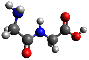
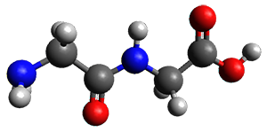
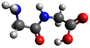
Continuing their application of laser ablation molecular beam Fourier transform microwave (LA-MB-FTMW) spectroscopy and computational chemistry to biochemical molecules (see these previous posts), the Alonso group reports on the structure of the glycine-glycine dipeptide 1.1 The microwave spectrum shows three different conformers. MP2/6-311++G(d,p) computations, the same method they have previously utilized for predicting geometries, revealed a number of different conformations. By matching the spectroscopic parameters obtained from the spectrum with those of the computed structures, they proposed the three conformations 1a1b, and 1c, shown in Figure 1.

1a

1b

1c
Figure 1. ωb97xd/6-31G(d) optimized structures of the three conformers of 1.
Note that the authors did not report their structures in their supporting materials(!) so I have optimized them.

The structures of conformers 1a and 1b are nearly planar. MP2 predicts a non-planar rotomer of 1a, which brings the carboxyl group out of plane, to be the lowest conformation in terms of electronic energy. With the M06-2x functional, this non-planar rotomer is about isoenergetic with 1a. With all computational levels 1a is the lowest in free energy. The barrier for rotation between the non-planar rotomer and 1a is very small, and this explains why it is not observed in the supersonic expansion.

References

1) Cabezas, C.; Varela, M.; Alonso, J. L., "The Structure of the Elusive Simplest Dipeptide Gly-Gly." Angew. Chem. Int. Ed. 2017, 56, 6420-6425, DOI: 10.1002/anie.201702425.

InChIs

1: InChI=1S/C4H8N2O3/c5-1-3(7)6-2-4(8)9/h1-2,5H2,(H,6,7)(H,8,9)
InChIKey=YMAWOPBAYDPSLA-UHFFFAOYSA-N


'
This work is licensed under a Creative Commons Attribution-NoDerivs 3.0 Unported License.

Tuesday, April 7, 2015

The Nucleoside Uridine Isolated in the Gas Phase

Peña, I.; Cabezas, C.; Alonso, J. L. Angew. Chem. Int. Ed. 2015, 54, 2991-2994
Contributed by Steven Bachrach.
Reposted from Computational Organic Chemistry with permission

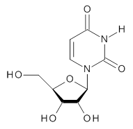
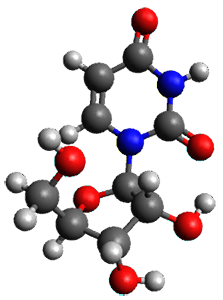
To advance our understanding of why ribose takes on the furanose form, rather than the pyranose form, in RNA, Alonso and co-workers have examined the structure of uridine 1 in the gas phase.1

1
Uridine is sensitive to temperature, and so the laser-ablation method long used by the Alonso group is ideal for examining uridine. The microwave spectrum is quite complicated due to the presence of many photofragments. Careful analysis lead to the identification of a number of lines and hyperfine structure that could be definitively assigned to uridine, leading to experimental values of the rotational constants and the diagonal elements of the 14N nuclear quadrupole coupling tensor for each nitrogen. These values are listed in Table 1.

Table 1. Experimental and calculated rotational constants (MHz), quadrupole coupling constants (MHz) and relative energy (kcal mol-1).


calculated


Expt.
anti/C2’-endo-g+
syn/C2’-endo-g+
anti/C3’-endo-g+
anti/C2’-endo-t
syn/C3’-endo-g+
A
885.98961
901.2
935.8
790.0
799.7
925.5
B
335.59622
340.6
308.4
352.6
330.6
300.4
C
270.11210
276.6
266.6
261.4
262.9
264.0
14N1χxx
1.540
1.50
1.82
1.48
1.46
1.82
14N1χyy
1.456
1.43
0.73
1.71
1.81
-0.72
14N1χzz
-2.996
-2.93
-2.56
-3.19
-3.27
-1.11
14N3χxx
1.719
1.74
2.03
1.78
1.62
1.98
14N3χyy
1.261
1.11
0.47
1.34
1.51
-0.75
14N3χzz
-2.979
-2.85
-2.50
-3.12
-3.13
-1.23
Rel E

0.0
1.10
1.90
2.00
2.15

In order to assign a 3-D structure to these experimental values, they examined the PES of uridine with molecular mechanics and semi-empirical methods, before reoptimizing the structure of the lowest 5 energy structures at MP2/6-311++G(d,p). Then, comparison of the resulting rotational constants and 14N nuclear quadrupole coupling constants of these computed structures (see Table 1) led to identification of the lowest energy structure (anti/C2’-endo-g+, see Figure 1) in best agreement with the experiment.Once again, the Alonso group has demonstrated the value of the synergy between experiment and computation in structure identification.

Figure 1. MP2/6-311++G(d,p) optimized structure of 1 (anti/C2’-endo-g+).


References

(1) Peña, I.; Cabezas, C.; Alonso, J. L. "The Nucleoside Uridine Isolated in the Gas Phase," Angew. Chem. Int. Ed. 201554, 2991-2994, DOI: 10.1002/anie.201412460.


Inchis:

1: Inchi=1S/C9H12N2O6/c12-3-4-6(14)7(15)8(17-4)11-2-1-5(13)10-9(11)16/h1-2,4,6-8,12,14-15H,3H2,(H,10,13,16)/t4-,6-,7-,8-/m1/s1
InChiKey=DRTQHJPVMGBUCF-XVFCMESISA-N


This work is licensed under a Creative Commons Attribution-NoDerivs 3.0 Unported License.

Tuesday, December 2, 2014

Tautomerism in Neutral Histidine

Bermúdez, C.; Mata, S.; Cabezas, C.; Alonso, J. L. Angew. Chem. Int. Ed. 2014, 53, 11015-11018
Contributed by Steven Bachrach.
Reposted from Computational Organic Chemistry with permission

The Alonso group has yet again (see these posts) determined the gas-phase structure of an important, biologically significant molecule using a combination of exquisite microwave spectroscopy and quantum computations. This time they examine the structure of histidine.1

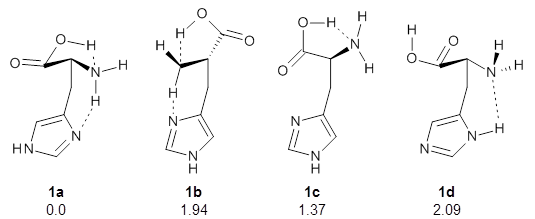
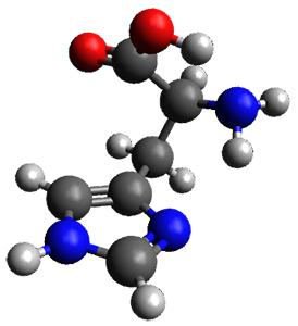
They optimized four conformations of histidine, as its neutral tautomer, at MP2/6-311++G(d,p). These are schematically drawn in Figure 1. Conformer 1a is the lowest in free energy, likely due to the two internal hydrogen bonds. Its structure is shown in Figure 2.

Figure 1. The four conformers of histidine. The relative free energy (MP2/6-311++G(d,p)) in kcal mol-1are also indicated.

Figure 2. MP2/6-311++G(d,p) optimized geometry of 1a.

The initial experimental rotation constants were only able to eliminate 1b from consideration. So they then determined the quadrupole coupling constants for the 14N nuclei. These values strongly implicated 1a as the only structure in the gas phase. The agreement between the experimental values and the computed values at MP2/6-311++G(d,p) was a concern, so they rotated the amine group to try to match the experimental values. This lead to a change in the NHCC dihedral value of -16° to -23° Reoptimization of the structure at MP2/cc-pVTZ led to a dihedral of -21° and overall excellent agreement between the experimental spectral parameters and the computed values.

It is somewhat disappointing the supporting materials does not include the structures of the other three isomers, nor the optimized geometry at MP2/cc-pVTZ.

References

1) Bermúdez, C.; Mata, S.; Cabezas, C.; Alonso, J. L. "Tautomerism in Neutral Histidine," Angew. Chem. Int. Ed. 201453, 11015-11018, DOI: 10.1002/anie.201405347.

InChIs

Histidine: InChI=1S/C6H9N3O2/c7-5(6(10)11)1-4-2-8-3-9-4/h2-3,5H,1,7H2,(H,8,9)(H,10,11)/t5-/m0/s1
InChIKey=HNDVDQJCIGZPNO-YFKPBYRVSA-N


This work is licensed under a Creative Commons Attribution-NoDerivs 3.0 Unported License.

Wednesday, February 19, 2014

How Sugars Pucker: Electronic Structure Calculations Map the Kinetic Landscape of Five Biologically Paramount Monosaccharides and Their Implications for Enzymatic Catalysis

Mayes, H. B.; Broadbelt, L. J.; Beckham, G. T. Journal of the American Chemical Society 2013, 136, 1008
Contributed by Steven Bachrach.
Reposted from Computational Organic Chemistry with permission

The conformational space of monosaccharides is amazingly complex. If we consider just the pyranose form, the ring can in principal exist as a chair, a half-chair, skew (or twist boat) and boat form, for a total of 38 puckering configurations. Layer on top of this the axial and equatorial positions of the hydroxyl and methylhydroxyl groups, and then the rotamers of these substituents, and one is faced with a dauntingly vast space. It is just this space that Beckham and co-workers1 take on for α- and β-glucose, β-xylose, β-mannose and β-acetylglucosamine.

For each sugar, and for each of the 38 puckering configurations, full rotamer scans for each of the substituents led to 27,702 conformations of each of the four monosaccharides, and 36,936 conformations of β-acetylglucosamine. This totals to over 123,000 geometry optimizations that were carried out at M06-2x/6-31G(d). Then taking the structures within 5 kcal mol-1 of the lowest energy structure for ­each pucker, they reoptimized at M06-2X/6-31+G(d,p). Pruning once again those structures that were above 5 kcal mol-1 of the minimum, they performed CCSD(T)/6-311+G(d,p)//B3LYP/6-311+G(2df,p) computations. What a tour de force!

The results of these conformational space surveys are not terribly exciting. The substituents do make a difference in dictating the most and least favorable structures and the activation barriers for interconversion of ring forms.

These PESs will be quite useful in understanding carbohydrate conformations and the role these may play in their chemistry. But the point of bringing this paper to your attention is the tremendously complex, detailed PES that is uncovered, representing the scale of what can be done with modern computers and modern algorithms.


References

(1) Mayes, H. B.; Broadbelt, L. J.; Beckham, G. T. "How Sugars Pucker: Electronic Structure Calculations Map the Kinetic Landscape of Five Biologically Paramount Monosaccharides and Their Implications for Enzymatic Catalysis," Journal of the American Chemical Society 2013136, 1008-1022, DOI:10.1021/ja410264d.


InChIs

α-glucose: InChI=1S/C6H12O6/c7-1-2-3(8)4(9)5(10)6(11)12-2/h2-11H,1H2/t2-,3-,4+,5-,6+/m1/s1
InChIKey: WQZGKKKJIJFFOK-DVKNGEFBSA-N
β-glucose: InChI=1S/C6H12O6/c7-1-2-3(8)4(9)5(10)6(11)12-2/h2-11H,1H2/t2-,3-,4+,5-,6-/m1/s1
InChIKey=WQZGKKKJIJFFOK-VFUOTHLCSA-N
β-xylose: InChI=1S/C5H10O5/c6-2-1-10-5(9)4(8)3(2)7/h2-9H,1H2/t2-,3+,4-,5-/m1/s1
InChIKey=SRBFZHDQGSBBOR-KKQCNMDGSA-N
β-mannose: InChI=1S/C6H12O6/c7-1-2-3(8)4(9)5(10)6(11)12-2/h2-11H,1H2/t2-,3-,4+,5+,6-/m1/s1
InChIKey=WQZGKKKJIJFFOK-RWOPYEJCSA-N
β-acetylglucosamine: InChI=1S/C8H15NO6/c1-3(11)9-5-7(13)6(12)4(2-10)15-8(5)14/h4-8,10,12-14H,2H2,1H3,(H,9,11)/t4-,5-,6-,7-,8-/m1/s1
InChIKey=OVRNDRQMDRJTHS-FMDGEEDCSA-N


This work is licensed under a Creative Commons Attribution-NoDerivs 3.0 Unported License.