Wednesday, September 30, 2015

Experimental Protein Structure Verification by Scoring with a Single, Unassigned NMR Spectrum


Contributed by Jan Jensen

Sometimes you read an article and think "wow, is it really that easy?"  This paper by Rienstra and co-workers is one of them. Protein structure determination based purely on chemical shifts is hard. Protein structure determination based purely on unassigned chemical shifts is even harder and I freely admit that I don't understand half of what is written in most papers on automated chemical shifts assignments.

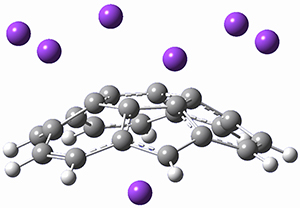
The method called, COMPASS, has 4 steps

1. An unassigned 2D $^{13}$C-$^{13}$C spectrum of aliphatic carbon atoms is measured experimentally for a given protein.

2. A standard MODELLER-based protocol is used to generate hundreds of candidate structures for the same protein.

3. SHIFTX2 is used to compute a 2D $^{13}$C-$^{13}$C spectrum for each candidate structure, i.e. all covalently bonded aliphatic carbons contribute a peak.

4. The measured spectrum is compared to each computed spectrum (based only a simple peak-closest peak distance criterion) and the candidate structure that results in the best match is predicted protein structure.

And it works:
We demonstrate COMPASS with experimental data from four proteins—GB1, ubiquitin, DsbA, and the extracellular domain of human tissue factor—and with reconstructed spectra from 11 additional proteins. For all these proteins, with molecular mass up to 25 kDa, COMPASS distinguished the correct fold, most often within 1.5 Å root-mean- square deviation of the reference structure.
Well, there is one protein (stR65) for which COMPASS didn't work as MODELLER did not produce any candidate structures whose C$\alpha$ RMSD was within 10 Å of the x-ray structure.  One cannot tell that the approach fails directly from the spectrum-comparison score but the authors show that, in case only, candidate structures with the five lowest spectrum-comparison scores differ wildly from each other.  So it looks like the COMPASS approach can also identify problem cases.

I was initially quite surprised that SHIFTX2 was sufficiently sensitive to changes in the protein structure for this to work but as the authors point out that "the scores depend not only on the C$\alpha$-C$\beta$ correlations, which report most strongly on secondary structure, but also on cross-peaks involving side-chain carbons, which report more strongly on the local environment."

It would of course be interesting to see if the spectrum-comparison score can be used as part of a hybrid energy function in a Monte-Carlo simulation to help guide the conformational sampling towards the correct structure.

I thank Kaare Teilum for bringing this paper to my attention


This work is licensed under a Creative Commons Attribution 4.0

Wednesday, September 23, 2015

Formation of Ground State Triplet Diradicals from Annulated Rosarin Derivatives by Triprotonation

Fukuzumi, S.; Ohkubo, K.; Ishida, M.; Preihs, C.; Chen, B.; Borden, W. T.; Kim, D.; Sessler, J. L.  J. Am. Chem. Soc. 2015, 137, 9780-9783
Contributed by Steven Bachrach.
Reposted from Computational Organic Chemistry with permission

What is the spin state of the ground state of an aromatic species? Can this be spin state be manipulated by charge? These questions are addressed by Borden, Kim, Sessler and coworkers1 for the hexaphyrin 1. B3LYP/6-31G(d) optimization of 1 shows it to be a ground state singlet. This structure is shown in Figure 1.

1

13+
Protonation of the three pyrrole nitrogens creates 13+, which has interesting frontier orbitals. The HOMO of 13+, of a1” symmetry, has nodes running through all six nitrogens. The next higher energy orbital, of a2” symmetry, has a small π-contribution on each nitrogen. Protonation will therefore have no effect on the energy of the a1” orbital, but the charge will stabilize the a2” orbital. This will lower the energy gap between the two orbitals, suggesting that a ground state triplet might be possible. The lowest singlet and triplet states of 13+ are also shown in Figure 1.

1

Singlet 13+

Triplet 13+
Figure 1. (U)B3LYP/6-31G(d) optimized structures of 1 and singlet and triplet 13+.

This spin state change upon protonation was experimentally verified by synthesis of two analogues of 1, shown below. The triprotonated versions of both are observed to have triplet character in their EPR spectrum.


References

(1) Fukuzumi, S.; Ohkubo, K.; Ishida, M.; Preihs, C.; Chen, B.; Borden, W. T.; Kim, D.; Sessler, J. L. "Formation of Ground State Triplet Diradicals from Annulated Rosarin Derivatives by Triprotonation," J. Am. Chem. Soc. 2015137, 9780-9783, DOI: 10.1021/jacs.5b05309.


InChIs

1: InChI=1S/C45H24N6/c1-2-8-29-28(7-1)34-16-22-13-24-18-36-30-9-3-4-10-31(30)38-20-26(50-43(38)42(36)48-24)15-27-21-39-33-12-6-5-11-32(33)37-19-25(49-44(37)45(39)51-27)14-23-17-35(29)41(47-23)40(34)46-22/h1-21,46,49-50H/b22-13-,23-14-,24-13-,25-14-,26-15-,27-15-
InChIKey=UMEHBSBBAUKCTH-UIJINECUSA-N


This work is licensed under a Creative Commons Attribution-NoDerivs 3.0 Unported License.

Thursday, September 17, 2015

Convex and Concave Encapsulation of Multiple Potassium Ions by Sumanenyl Anions

Spisak, S. N.; Wei, Z.; O’Neil, N. J.; Rogachev, A. Y.; Amaya, T.; Hirao, T.; Petrukhina, M. A. J. Am. Chem. Soc. 2015, 137, 9768-9771
Contributed by Steven Bachrach.
Reposted from Computational Organic Chemistry with permission

Spisak, et al. treated sumanene 1 with excess potassium in THF.1

They obtained an interesting structure, characterized by x-ray crystallography: a mixture of the dianion and trianion of 1 (well these are really conjugate di- and tribases of 1, but we’ll call them di- and trianions for simplicity’s sake). A fragment of the x-ray structure is shown in Figure 1, showing that there is one potassium cation on the concave face and six potassium ions on the convex face.

Figure 1. X-ray structure of 1 surrounded by six K+ ions on the convex face and one K+ on the concave face.

To help understand this structure, they performed RIJCOSX-PBE0/cc-pVTZ computations on the mono-, di-, and trianion of 1. The structure of 1 (which I optimized at ωB97X-D/6-311G(d)) and the trianion are displayed in Figure 2. The molecular electrostatic potential of the trianion shows highly negative regions in the 5-member ring regions, symmetrically distributed and prime for coordination with 6 cations.

1

trianion of 1
Figure 2. Optimized structure of 1 and its trianion.


References

(1) Spisak, S. N.; Wei, Z.; O’Neil, N. J.; Rogachev, A. Y.; Amaya, T.; Hirao, T.; Petrukhina, M. A. "Convex and Concave Encapsulation of Multiple Potassium Ions by Sumanenyl Anions," J. Am. Chem. Soc. 2015137, 9768-9771, DOI: 10.1021/jacs.5b06662.


InChIs

1: InChI=1S/C21H12/c1-2-11-8-13-5-6-15-9-14-4-3-12-7-10(1)16-17(11)19(13)21(15)20(14)18(12)16/h1-6H,7-9H2
InChIKey=WOYKPMSXBVTRKZ-UHFFFAOYSA-N
Trianion of 1: InChI=1S/C21H9/c1-2-11-8-13-5-6-15-9-14-4-3-12-7-10(1)16-17(11)19(13)21(15)20(14)18(12)16/h1-5,7-8H,9H2/q-3
InChIKey=IHJVIPHOCKVJDZ-UHFFFAOYSA-N


This work is licensed under a Creative Commons Attribution-NoDerivs 3.0 Unported License.