Wednesday, August 29, 2012

Effect of Hydrogen Bonds on pKa Values: Importance of Networking

Shokri, A.; Abedin, A.; Fattahi, A.; Kass, S. R.  J. Am. Chem. Soc. 2012, 134, 10646 (Paywall)
Contributed by Steven Bachrach.
Reposted from Computational Organic Chemistry with permission

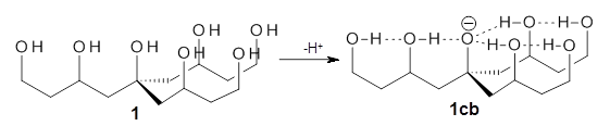
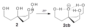
Can a hydrogen bonding network affect acidity? Kass has examined the polyol 1 whose conjugate base1cb can potentially be stabilized by a large hydrogen bonding network.1 Kass had previously found a significant acidy enhancement in comparing t-butanol (ΔG(deprotonation) = 369.2 kcal mol-1) with that of 2 (ΔG(deprotonation) = 334.4 kcal mol-1).2

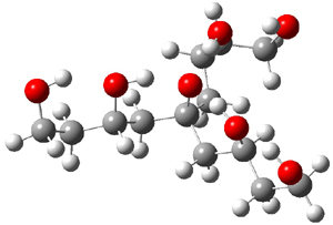
Table 1 lists the computed and experimental free energies of deprotonation of 1. The experimental values are computed at M06-2x/maug-cc-pVT(+d)Z. The structure of 1cb is drawn in Figure 1.

Table 1. Computed and experimental free energies (kcal mol-1 of deprotonation of some alcohols.

MO6-2x
Expt
t-butanol
368.6
369.3
2
335.0
334.4
1
320.2
313.5
The difference in the acidity of t-butanol and 2, some 30 kcal mol-1, reflects the stability afforded by three intramolecular hydrogen bonds to the oxyanion. In going from 2cb to 1cb, each of the hydroxyl groups that donate to the oxyanion act as the acceptor of a hydrogen bond from the more removed hydroxyl groups. There is in effect a first and second layer of hydrogen bond network in 1cb. These secondary hydrogen bonds lead to further stabilization of the anion, as reflected in the diminished DPE of 1 over 2: 320.2 vs. 335.0 kcal mol-1. Note that this secondary layer does not stabilize the anion to the same degree as the primary layer, but nonetheless its effect is large and quite striking.

1cb
Figure 1. M06-2x/maug-cc-pVT(+d)Z optimized structure of 1cb.

Even in solution these more remote hydrogen bonds can stabilize the anion. So, using the CPCM approach and modeling DMSO, 2 is predicted have a pKa that is 15 units below that of t-butanol, and 1is predicted to be 3 pKa units more acidic than 2. Experiments verify this prediction with the pKas of 16.1 for 2 and 11.4 for 1.

References

(1) Shokri, A.; Abedin, A.; Fattahi, A.; Kass, S. R. "Effect of Hydrogen Bonds on pKa Values: Importance of Networking," J. Am. Chem. Soc. 2012134, 10646-10650, DOI: 10.1021/ja3037349
(2) Tian, Z.; Fattahi, A.; Lis, L.; Kass, S. R. "Single-Centered Hydrogen-Bonded Enhanced Acidity (SHEA) Acids: A New Class of Brønsted Acids," J. Am. Chem. Soc. 2009131, 16984-16988, DOI:10.1021/ja9075106

InChIs

1: InChI=1S/C13H28O7/c14-4-1-10(17)7-13(20,8-11(18)2-5-15)9-12(19)3-6-16/h10-12,14-20H,1-9H2
InChIKey=HGTVPOTWAYDRSM-UHFFFAOYSA-N
1cb: InChI=1S/C13H27O7/c14-4-1-10(17)7-13(20,8-11(18)2-5-15)9-12(19)3-6-16/h10-12,14-19H,1-9H2/q-1
InChIKey=UQPPTNIHRICITD-UHFFFAOYSA-N
2: InChI=1S/C7H16O4/c8-4-1-7(11,2-5-9)3-6-10/h8-11H,1-6H2
InChIKey=FAQWYKIIWYVDPQ-UHFFFAOYSA-N
2cb: InChI=1S/C7H15O4/c8-4-1-7(11,2-5-9)3-6-10/h8-10H,1-6H2/q-1
InChIKey=WSCPTRIAWKZJFZ-UHFFFAOYSA-N


Thursday, August 23, 2012

Refinement of protein structure homology models via long, all-atom molecular dynamics simulations

Alpan Raval, Stefano Piana, Michael P. Eastwood, Ron O. Dror, and David E. Shaw, Proteins 2012, 80, 2071-2079 (Paywall)
Contributed by Victor Guallar


Many theoretical chemists work routinely on biological systems and, in particular, on proteins. While it might not be their main interest, predicting the conformational sampling associated to these systems is certainly a concern.  Those who have been around for a while have seen how the necessary conformational sampling has moved from few picoseconds to hundreds of nanoseconds and even microseconds (while I do not agree, molecular dynamics has almost the exclusivity as a sampling technique). Clearly the latest development of special-purpose computers, such as the remarkable effort from the D. E. Shaw Research group, together with the development of molecular dynamics for graphical processing units, have contributed to this time expansion. Along these advances we surely had the following questions: are the force fields up to it?, how meaningful are these long molecular dynamics simulations?

The Shaw group has probably already answered these questions for us. In a comprehensive study1 they produce at least a hundred microseconds simulation for 24 proteins used in recent CASP competitions. They frame their study under the capabilities of molecular dynamics (and force fields) in refining homology models. Thus, for each system they produce a trajectory from both an initial homology model and from the native X-ray structure (or NMR). This study followed a previous one where the simulations were capable of accurately reproducing the native state on several fast-folders. The results this time, however, are quite surprising and even worrisome. For most of the systems the structures drift away from the native state. Furthermore, this drift occurs even when starting from the native state. Overall the results indicate that for most systems the force field minimum is not consistent with the X-ray or NMR experimental structures. While the authors only used two force fields (considered to be the best ones), they conclude that most likely this is a limitation for all available force fields.

The authors obtain better results when imposing constraints to the simulation (limiting the drift away from the native structure). Thus, one can conclude from this work that brute force molecular dynamics simulations are still far away from being accurate. Obviously similar conclusion could be applied to other sampling techniques using the same force fields (for example Monte Carlo techniques). While we wait for better force fields (maybe polarizable ones such AMOEBA?), we probably should use molecular dynamics as a local exploration rather than to predict novel conformations, or to score significantly different ones. Of course these limitations might not apply to those systems with a strong preference for a state, such as fast-folder proteins.

References
Alpan Raval, Stefano Piana, Michael P. Eastwood,Ron O. Dror, and David E. ShawProteins 2012, 80, 2071-2079 

Saturday, August 11, 2012

Transition states of native and drug-resistant HIV-1 protease are the same


What is the rate determining step?
The general features of the HIV-1 protease mechanism appear to be 1) nucleophilic attack on the amide carbonyl group by a water molecule to form a gem-diol intermediate, 2) protonation of the amide nitrogen, and 3) cleavage of the amide C-N bond.

As pointed out in the introduction, computational studies such as Okimoto et al. and. Piana et al. both  seem to indicate that Step 2 is the rate limiting step.

However, a closer look at these studies suggest that the issue is not well settled.  Okimoto et al.'s (small gas phase) model suggests that the gem-diol intermediate is more stable than the enzyme-substrate complex and computed the barrier of the second step relative to this intermediate.  Piana et al.'s (QM/MM) model suggests that the gem-diol intermediate is less stable than the enzyme-substrate complex and computed the barrier of the second step relative to this enzyme substrate.

If one switches the reference state in both studies Okimoto et al.'s model would predict the first two steps to have essentially the same barrier, while Piana et al.'s model would predict the first step to be rate determining.  Furthermore, the primary $^{15}$N isotope effect computed by Piana et al. (0.97$\pm$0.2) is larger than the experimental value obtained by Rodrigez et al. (0.995$\pm$0.002) (large isotope effects mean significant deviations from 1).

Kinetic isotope effects confirm the rate limiting step and the transition state structure
Schramm and co-workers have used labelled substrates to measure primary $^{14}$C and $^{15}$N and secondary $^3$H and $^{18}$O isotope effects.  Furthermore, they used small gas phase models to compute the corresponding isotope effects for all five stationary points at the ONIOM (M06-2X/6-31+G**:am1) level of theory.

Overall the measured isotope effects best match the computed values for the transition state for Step 2, which is therefore likely to be the rate determining step.  This isotope effect was computed relative to the enzyme-substrate complex; it would be interesting to recompute this value relative to the gem-diol intermediate, which is sufficiently stable to be observed experimentally at low-pH conditions (Das et al.)

Transition states and drug design
Very similar isotope effects was also measured for a mutant (I84V) which has displayed resistance to all nine FDA approved-inhibitors, indicating that the transition state structure in this mutant is quite similar to that of the wild-type.  Thus, transition state-mimics would likely inhibit both forms of the enzyme and may lead to new inhibitors that are less prone to resistance.

Acknowledgement: Thanks to Luca De Vico for alerting me to this paper

Creative Commons License
This work is licensed under a Creative Commons Attribution 3.0 Unported License.