Tuesday, January 7, 2014

Equatorenes: Synthesis and Properties of Chiral Naphthalene, Phenanthrene, Chrysene, and Pyrene Possessing Bis(1-adamantyl) Groups at the Peri-position

Yamamoto, K.; Oyamada, N.; Xia, S.; Kobayashi, Y.; Yamaguchi, M.; Maeda, H.; Nishihara, H.; Uchimaru, T.; Kwon, E. J. Am. Chem. Soc. 2013,135, 16526
Contributed by Steven Bachrach.
Reposted from Computational Organic Chemistry with permission

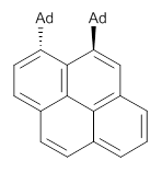
Naphthalene, phenanthrene and pyrene are all planar aromatic compounds. How can substituted version be chiral, with the chirality present in the aromatic portion of the molecule? The answer is provided by Yamaguchi and Kwon.1 They prepared peri-substituted analogues with the bulky adamantly group as the substituents. This bulky requires one adamantyl group to be position above the aromatic plane and the other below the plane, as in 1 and 2.
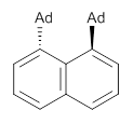
1

2
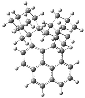
These molecules and two other examples were prepared in their optically pure form. B3LYP/6-31G(d) computations were performed on both of these structures (shown in Figure 1), but computations are a minor component of the work. These structures do show the out-of-plane distortions at C1 and C8, also apparent in the crystal structures. Computations of naphthalene and 1,8-dimethylnaphthalene show a planar naphthalene backbone, but i­-propyl substitution does force the substituents out of plane.
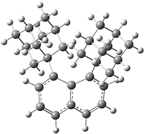
1

2
Figure 1. B3LYP/6-31G(d) optimized structures of 1 and 2.

These types of systems continue to subject the notion of “aromaticity” to serious scrutiny.


References

(1) Yamamoto, K.; Oyamada, N.; Xia, S.; Kobayashi, Y.; Yamaguchi, M.; Maeda, H.; Nishihara, H.; Uchimaru, T.; Kwon, E. "Equatorenes: Synthesis and Properties of Chiral Naphthalene, Phenanthrene, Chrysene, and Pyrene Possessing Bis(1-adamantyl) Groups at the Peri-position," J. Am. Chem. Soc. 2013,135, 16526-16532, DOI: 10.1021/ja407800e.


InChIs

1: InChI=1S/C30H36/c1-3-25-4-2-6-27(30-16-22-10-23(17-30)12-24(11-22)18-30)28(25)26(5-1)29-13-19-7-20(14-29)9-21(8-19)15-29/h1-6,19-24H,7-18H2
InChIKey=QNPJKZPPLCPHSS-UHFFFAOYSA-N
2: InChI=1S/C36H38/c1-2-27-4-5-28-6-7-30(35-15-21-8-22(16-35)10-23(9-21)17-35)34-31(14-29(3-1)32(27)33(28)34)36-18-24-11-25(19-36)13-26(12-24)20-36/h1-7,14,21-26H,8-13,15-20H2
InChIKey=DKRBDGNWYTWNHL-UHFFFAOYSA-N


This work is licensed under a Creative Commons Attribution-NoDerivs 3.0 Unported License.

Saturday, January 4, 2014

Electron correlation, zero-point vibrational and temperature effects in Nuclear Magnetic Resonance spectroscopy

A.M. Teale, O.B. Lutnæs, T. Helgaker, D.J. Tozer, J. Gauss, Journal of Chemical Physics 2013, 138, 024111 (Pay-wall) and
J. Kaminský, M. Buděšínský, S. Taubert, P. Bouřa, M. Straka, Physical Chemistry Chemical Physics 2013, 15, 9223-9230 (Open Access)
Contributed by Marcel Swart

Last year has seen two important contributions in the field of determination of NMR chemical shifts by theoretical chemistry. More and more it is recognized that the computational prediction of 1H and 13C chemical shifts is a useful tool for natural product, mechanistic, and synthetic organic chemistry.[1] There are however doubts about how accurate these results are, and if any chemically relevant conclusions can be drawn from them.

The first paper[2] compares the chemical shifts as obtained by both density functional theory and wavefunction theory (RHF, CCSD, CCSD(T), extrapolated) for a total of 28 molecules (for which previously already rotational g-tensors and magnetizabilities were computed[3]). The authors also included zero-point vibrational effects on the computed chemical shieldings, and used extrapolation techniques to estimate uncertainties related to basis-set incompleteness[4]. First, the authors established an accurate benchmark set of data, for which the accuracy was established by comparison with experimental data (including zero-point vibrational corrections). They found good agreement between CCSD(T)/aug-cc-pCVQZ and experiment (empirical equilibrium values), with a mean-absolute-error of 2.9 ppm for the chemical shieldings. Afterwards, these reference data were used to compare how well a variety of density functionals was able to reproduce them, with a sobering conclusion: "None of the existing approximate functionals provide an accuracy competitive with that provided by CCSD or CCSD(T) theory".[2] The best performing functional (as shown before) was KT2 with a mean-absolute-error compared to the CCSD(T) data (both with the same aug-cc-pCVQZ basis set) of 10.2 ppm.

The second paper[5] has a completely different approach, and deals with the characterization of fullerenes. For this purpose, computational chemistry might be used, but one again should be sure about the methods used. The authors used quantum vibrational averaging, a dielectric continuum model for the solvent (CPCM, 1,1,2-trichloroethane), classical (MM3) and first-principle (BP86/def-SVP) molecular dynamics simulations, and did experiments. These authors used a different set of density functionals, and found the best results for wB97X-D/IGLO-III (a root-mean-square deviation compared to experiment of only 0.4 ppm). However, surprisingly, in the analysis of the dynamical part they used either BP86 or BHandHLYP for the NMR chemical shifts, even though these showed larger RMSD values of respectively 1.7 and 0.8 ppm. Moreover, big differences were found in the chemical shifts from the snapshots of the 1 ns classical MD, and those of the 1.2 ps first-principles MD. For the five different atom types these differences were found in the range 1.8-7.9 ppm.[6]

References and notes
[1] M.W. Lodewyk, M.R. Siebert, D.J. Tantillo, Chem. Rev. 2012, 112, 1839-1862 [DOI 10.1021/cr200106v]
[2] A.M. Teale, O.B. Lutnæs, T. Helgaker, D.J. Tozer, J. Gauss, J. Chem. Phys. 2013, 138, 024111 [DOI 10.1063/1.4773016]
[3] O. B. Lutnæs, A. M. Teale, T. Helgaker, D. J. Tozer, K. Ruud, J. Gauss, J. Chem. Phys. 2009, 131, 144104 [DOI 10.1063/1.3242081]
[4] These formulas have been developed for energies; hence, their use for the direct extrapolation of molecular properties is less well founded, and indeed can not be used with a two-point extrapolation for prediction of the basis set limiting value.
[5] J. Kaminský, M. Buděšínský, S. Taubert, P. Bouřa, M. Straka, Phys. Chem. Chem. Phys. 2013, 15, 9223-9230 [DOI 10.1039/C3CP50657F]
[6] Strangely enough, while the CPCM solvent model only had a modest effect on the chemical shifts of 0.2-0.3 ppm, the authors showed that first-principles MD (FPMD) simulations including solvent effects (COSMO) led to drastically different results for the chemical shifts of snapshots (0.1-4.1 ppm) from those from FPMD without them.

Creative Commons License
This work is licensed under a Creative Commons Attribution 3.0 Unported License. 

Friday, January 3, 2014

CP and BSSE

Contributed by Frank Jensen

A couple of recent papers illustrate the problems related to using the CP correction for reducing BSSE:

Ł. M. Mentel and E. J. Baerends, JCTC ASAP, DOI: 10.1021/ct400990u
Report that for He-He and Be-Be interaction potentials, the CP correction can be in the wrong direction. The cause is apparently that the basis sets are optimized for the atoms, and thus biased against the complex. The uncorrected interaction energy is thus underestimated and adding the CP correction further underbinds the complex.

Lori A. Burns, Michael S. Marshall, and C. David Sherrill, JCTC ASAP, DOI: 10.1021/ct400149j
Perform a benchmark study using MP2 and CCSD(T) with and without CP corrections, or the average, combined with basis set extrapolation for the A24 benchmark systems + a few extras. Their conclusion is that whether to use the CP, half the CP or no CP depend on the system, method and basis set. Their (weak) recommendation is to use half the CP correction for basis sets of aDZP or aTZP quality to 'avoid the worst errors incurred by either method', and the full CP for larger basis sets and extrapolations.

The latter study most likely has components of the first: A given (fixed) basis set will be (slightly) non-optimum for each fragment and the complex, but which fragment/complex that it is least optimum for will depend on the system and geometry. The inherent basis set over/under-binding of the complex will be modulated by the overbinding by the BSSE. Adding the CP estimate of the BSSE can then lead to either improvement or deterioration of the final binding energy. Noting that all three effects are small, the inherent basis set error can have either sign, the BSSE is always negative and the CP correction is always positive, the combined effect will have significant 'random' errors compared to the exact result, which in magnitude also is small.

Thursday, January 2, 2014

Microscopic Insights into the NMR Relaxation-Based Protein Conformational Entropy Meter

Vignesh Kasinath, Kim A. Sharp, and A. Joshua Wand Journal of the American Chemical Society 2013, 135, 15092
Contributed by +Jan Jensen

Order parameters measured by NMR report on the local fluctuations of protein structures and should therefore be related to entropy. This study uses molecular dynamics (MD) simulations to obtain a quantitative relationship between conformational side chain entropy ($S_{sc}$) and Lipari-Szabo methyl group squared generalized order parameters ($O^2$).

First the authors demonstrate that MD simulations can reproduce experimentally measured $O^2$ values for 7 different proteins, with an $R^2$ of 0.92.

Second the authors demonstrate a linear correlation between the total $S_{sc}$ and computed $O^2$ values, both for methyl containing side chains ($R^2$ = 0.90) and for all side chains ($R^2$ = 0.91).

Based on these finding the authors re-analyzed data from two previously published studies, and extracted a similarly quantitative linear correlation ($R^2$ = 0.95) between the molecular entropy change ($\Delta S_{tot}-\Delta S_{solv}$) and the measured change in $O^2$ values for protein-protein and protein-DNA binding.  This suggest that both entropy changes are dominated by the changes in conformational side-chain entropy.

The method looks like a very powerful tool for obtaining detailed quantitative structural understanding of entropy changes in biomolecular processes.

Wednesday, January 1, 2014

Popular highlights of 2013

Computational Chemistry Highlights received 28,762 pageviews in 2013.

A total of 48 highlights were published in 2013 and the five most viewed highlights are

1. Are Protein Force Fields Getting Better? A Systematic Benchmark on 524 Diverse NMR Measurements highlighted by Ric Baron in March

2. Will molecular dynamics simulations of proteins ever reach equilibrium? highlighted by Gerald Monard in July

3. Molecularspace.org highlighted by Jan Jensen in June

4. A geometrical correction for the inter- and intra-molecular basis set superposition error in Hartree-Fock and density functional theory calculations for large systems highlighted by Steven Bachrach in January

5. Intrinsic Atomic Orbitals: An Unbiased Bridge between Quantum Theory and Chemical Concepts highlighted by Grant Hill in October

However, the most viewed post in 2013 was Chemical Networks (Triple header!) highlighted by Rob Paton in July 2012.

The most popular entries within the last 30 days can as always be found on the right hand side of this page.

The are many ways to stay updated on the latest CCH highlights and CCH currently has 119 subscribers on Feedly, 130 twitter followers, 476 Google+ followers, 43 Facebook followers, and 90 followers on Linkedin.

Creative Commons License
This work is licensed under a Creative Commons Attribution 4.0 International License.

Wednesday, December 18, 2013

Six Pyranoside Forms of Free 2-Deoxy-D-ribose

Peña, I.; Cocinero, E. J.; Cabezas, C.; Lesarri, A.; Mata, S.; Écija, P.; Daly, A. M.; Cimas, Á.; Bermúdez, C.; Basterretxea, F. J.; Blanco, S.; Fernández, J. A.; López, J. C.; Castaño, F.; Alonso, J. L.  Angew. Chem. Int. Ed. 2013, 52, 11840-11845
Contributed by Steven Bachrach.
Reposted from Computational Organic Chemistry with permission


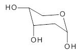
2-deoxyribose 1 is undoubtedly one of the most important sugars as it is incorporated into the backbone of DNA. The conformational landscape of 1 is complicated: it can exist as an open chain, as a five-member ring (furanose), or a six-member ring (pyranose), and intramolecular hydrogen bonding can occur. This internal hydrogen bonding is in competition with hydrogen bonding to water in aqueous solution. Unraveling all this is of great interest in predicting structures of this and a whole host of sugar and sugar containing-molecules.

1
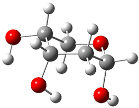
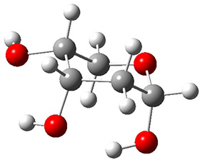
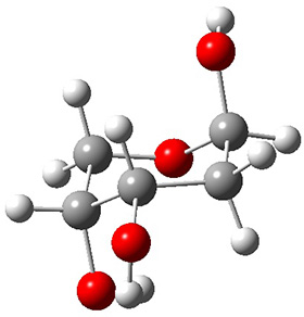
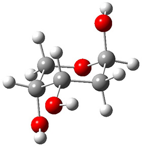
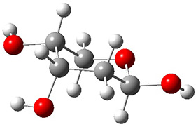
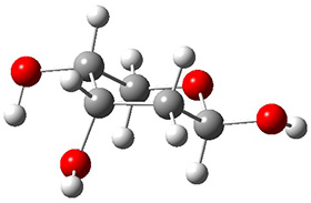
In order to get a firm starting point, the gas phase structures of the low energy conformers of 1 would constitute a great set of structures to use as a benchmark for gauging force fields and computational methods. Cocinero and Alonso1 have performed a laser ablation molecular beam Fourier transform microwave (LA-MB-FTMW) experiment (see these posts for other studies using this technique) on 1 and identified the experimental conformations by comparison to structures obtained at MP2/6-311++G(d,p). Unfortunately the authors do not include these structures in their supporting materials, so I have optimized the low energy conformers of 1 at ωB97X-D/6-31G(d) and they are shown in Figure 1.

1a (0.0)

1b (4.7)

1c (3.3)

1d (5.6)

1e (8.9)

1f (9.4)
Figure 1. ωB97X-D/6-31G(d) optimized structures of the six lowest energy conformers of 1. Relative free energy in kJ mol-1.

The computed spectroscopic parameters were used to identify the structures responsible for the six different ribose conformers observed in the microwave experiment. To give a sense of the agreement between the computed and experimental parameters, I show these values for the two lowest energy conformers in Table 1.

Table 1. MP2/6-311++G(d,p) computed and observed spectroscopic parameters for the two lowest energy conformers of 1.

1a
1c

Expt
Calc
Expt
Calc
A(MHz)
2484.4138
2492
2437.8239
2447
B (MHz)
1517.7653
1533
1510.7283
1527
C (MHz)
1238.9958
1250
1144.9804
1158
ΔG (kJ mol-1)

0.0

3.3
This is yet another excellent example of the symbiotic relationship between experiment and computation in structure identification.


References

(1) Peña, I.; Cocinero, E. J.; Cabezas, C.; Lesarri, A.; Mata, S.; Écija, P.; Daly, A. M.; Cimas, Á.; Bermúdez, C.; Basterretxea, F. J.; Blanco, S.; Fernández, J. A.; López, J. C.; Castaño, F.; Alonso, J. L. "Six Pyranoside Forms of Free 2-Deoxy-D-ribose," Angew. Chem. Int. Ed. 2013, 52, 11840-11845, DOI:10.1002/anie.201305589.


InChIs

1a: InChI=1S/C5H10O4/c6-3-1-5(8)9-2-4(3)7/h3-8H,1-2H2/t3-,4+,5-/m0/s1
InChIKey=ZVQAVWAHRUNNPG-LMVFSUKVSA-N


Creative Commons License
This work is licensed under a Creative Commons Attribution-NoDerivs 3.0 Unported License.

Tuesday, December 10, 2013

Computation-aided structure determination

(1) Jahn, M. K.; Dewald, D.; Vallejo-López, M.; Cocinero, E. J.; Lesarri, A.; Grabow, J.-U. "Rotational Spectra of Bicyclic Decanes: The Trans Conformation of (-)-Lupinine," J. Phys. Chem. A 2013
(2) Shepherd, D. J.; Broadwith, P. A.; Dyson, B. S.; Paton, R. S.; Burton, J. W. "Structure Reassignment of Laurefurenynes A and B by Computation and Total Synthesis," Chem. Eur. J. 2013, 19, 12644-12648
Contributed by Steven Bachrach.
Reposted from Computational Organic Chemistry with permission

I have not discussed any papers that utilize computations to confirm chemical structure in a while, so here are two recent examples.

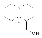
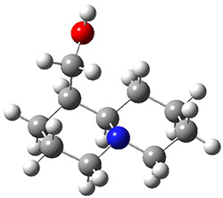
Grabow has utilized MP2 and M06-2x computations to confirm the lowest energy conformation of (-)-lupinine 1.1 The interesting structural aspect of this compound is the possibility of an intramolecular hydrogen bond linking the hydroxyl group with the amine.

1
Using molecular mechanics, the authors identified 57 structures within 50 kJ mol-1 of each other. These geometries were reoptimized at MP2/6-311++G(d,p) and M06-2x/6-311++G(d,p).

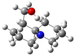
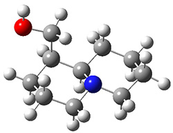
The lowest energy structures had the expected trans ring fusion, with a trans relationship between the hydrogen on the bridgehead carbon (C9) and the hydroxymethyl group. This corresponds to either the (R,R) or (S,S) isomer. The three lowest energy structures are shown in Figure 1. Unfortunately, the geometry for the lowest energy isomer provided in the Supporting Materials is wrong, and the authors did not supply the geometries of the other isomers. This situation is unacceptable! Reviewers and editors must do a better job in policing the Supporting Materials; there is no excuse for not including all of the optimized structures, and better yet, in a more usable format that what has been done here. I have reoptimized these structures at M06-2x/6-31G(d). The lowest energy conformer 1a does possess the expected internal hydrogen bond.

1a
(0.0)

1b
(10.4)

12
(11.5)
Figure 1. M06-2x/6-31G(d) optimized structures of the three lowest energy conformers of 1, with relative free energies in kJ mol-1.

Table 1 provides a comparison of the MP2 computed values of important structural parameters along with the experimental values obtained from a microwave experiment. The agreement with the computed values for 1a provides strong evidence that this is the structure of (-)-lupinine.

Table 1. Comparison of MP2 and experimental structural parameters of 1.a

Expt. (1)
MP2 (1a)
A
1414.126
1425.8
B
811.672
815.1
C
671.530
677.1
ΔJ
0.0255
0.023
ΔJK
0.0639
0.065
ΔK
0.0037
0.0022
χaa
1.9973
2.0
χbb
1.062
1.1
χcc
-3.059
-3.1
aRotational constants (A, B, C) in MHz, centrifugal distortion constants (ΔJ, ΔJK, ΔK) in kHz, and nuclear quadrupole coupling tensor elements (χaa, χbb, χcc) in MHz.

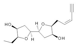
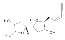
The second study utilizes computed NMR chemical shifts to discriminate potential diastereomeric structures. Laurefurenyne A was first assigned the structure 2 based on 1D and 2D NMR experiments. However, based on potential biochemical analogy to other compounds, Paton and Burton2 had doubts about this structure. In addition to synthesizing the natural material, they performed an extensive computational study of the chemical shifts of the diastereomers. For each of the 32 possible diastereomers, they performed a Monte Carlo search of the conformational space using molecular mechanics. The structures of all isomers within 10 kJ mol-1 of the lowest energy structure were reoptimized at ωB97X-D/6-31G(d) with PCM (CHCl3) and chemical shifts obtained at mPW1PW91/6-311G(d,p). Final chemical shifts were obtained using a Boltzmann weighting. The computed values for 2were quite off from the experimental values, with a mean unsigned error of 1.5 ppm. A better assessment was provided with the DP4 method, which indicated that 3 has the highest probability of being the correct structure, a structure consistent with the likely biosynthetic pathway.

2

3


References

(1) Jahn, M. K.; Dewald, D.; Vallejo-López, M.; Cocinero, E. J.; Lesarri, A.; Grabow, J.-U. "Rotational Spectra of Bicyclic Decanes: The Trans Conformation of (-)-Lupinine," J. Phys. Chem. A 2013, DOI:10.1021/jp407671m.
(2) Shepherd, D. J.; Broadwith, P. A.; Dyson, B. S.; Paton, R. S.; Burton, J. W. "Structure Reassignment of Laurefurenynes A and B by Computation and Total Synthesis," Chem. Eur. J. 2013, 19, 12644-12648, DOI: 10.1002/chem.201302349.


InChIs

(-)-Lupinine 1: InChI=1S/C11H21NO/c1-11-6-2-3-7-12(11)8-4-5-10(11)9-13/h10,13H,2-9H2,1H3/t10-,11+/m0/s1
InChIKey=WVDUAOYJLFVEMW-WDEREUQCSA-N
Laurefurenyne A 3: InChI=1S/C14H20O4.C2H6/c1-3-4-5-6-12-11(16)8-14(18-12)13-7-10(15)9(2)17-13;1-2/h1,4-5,9-16H,6-8H2,2H3;1-2H3/b5-4-;/t9-,10-,11-,12+,13-,14+;/m1./s1
InChIKey=ZYESDGGYHKCHMJ-SKFGUVSTSA-N